
ECG Made Easy - Sinus Node Dysfunction Explained Clearly
Sinus arrest/pause and SA exit blocks explained in 4 min - It's mind-boggling how easy it is!
This lesson includes an animated video lecture, downloadable images, quiz questions and a PDF
The heart electrical signals are initiated in its natural pacemaker - the sinoatrial node, or SA node, and travel through the atria to reach the atrioventricular node, or AV node. The AV node passes the signals onto the bundle of His, which then splits into two branches that conduct the impulses to the two ventricles.
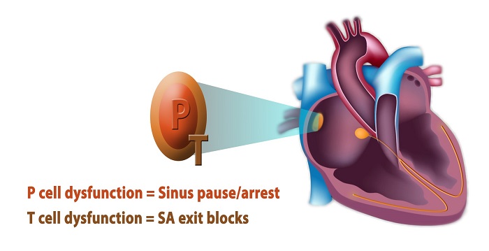
The SA node is composed of two major cell types:
- P, for pacemaker, cells generate electrical impulses;
- and T, for transitional, cells transmit these signals to the right atrium and subsequently to the rest of the cardiac conduction system.
Sinus node dysfunction occurs when any of these cells cease to function properly:
- failure to produce electrical impulses by P cells leads to sinus pause or sinus arrest,
- while delay or failure of signal transmission by T cells results in SA exit blocks.
On an ECG, sinus pause or arrest can be seen as a brief absence of P waves, which can last for seconds to minutes. In most cases, a downstream pacemaker in the atria, atrioventricular junction, or ventricles, will take over the pacing function, producing so-called escape beats or rhythms, and thus preserving heart rate and function until the SA node recovers and fires again; but long pauses may cause dizziness, fainting and possibly cardiac arrest.
Subscribe to one of the courses below to continue!
This content is available within the following courses:

Our Signature Animated Videos on Electrocardiography: 25 animations, plus downloadable PDFs, downloadable images, and quizzes.

Our Signature Animated Videos on Electrocardiography: 25 animations, plus downloadable PDFs, downloadable images, and quizzes.