
Action Potential in Neurons Explained Clearly
One of the Most Difficult Topics of Neurobiology Explained Clearly in 6 min - A Mini-Lecture by Alila Medical Media.
This lesson includes an animated video lecture, downloadable images, quiz questions and a PDF
Neurons communicate with each other through their dendrites and axon. Generally, incoming signals are received at dendrites, while outgoing signal travels along the axon to the nerve terminal. In order to achieve rapid communication over its long axon, the neuron sends electrical signals, known as nerve impulses, or action potentials, from the cell body to the nerve terminal.
An action potential is essentially a brief reversal of electric polarity of the cell membrane.
Cells are polarized, meaning there is an electrical voltage across the cell membrane. In a resting neuron, the typical voltage, known as the resting membrane potential, is about -70mV. The negative value means the cell is more negative on the inside.
At this resting state, there are concentration gradients of sodium and potassium across the membrane: more sodium outside the cell and more potassium inside the cell. These gradients are maintained by the sodium-potassium pump which constantly brings potassium in and pumps sodium out of the cell.
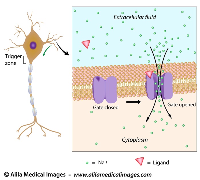
A neuron is typically stimulated at the dendrites and the signals spread through the soma. Excitatory signals at dendrites open ligand-gated sodium channels and allow sodium to flow into the cell. This neutralizes some of the negative charge inside the cell and makes the membrane voltage less negative. This is known as depolarization as the cell membrane becomes less polarized.
The influx of sodium diffuses inside the neuron and produces a current that travels toward the axon hillock. If the summation of all input signals is excitatory and is strong enough when it reaches the axon hillock, an action potential is generated and travels down the axon to the nerve terminal.
The axon hillock is known as the cell’s “trigger zone” as this is where action potentials usually start. This is because action potentials are produced by voltage-gated ion channels that are most concentrated at the axon hillock.
Voltage-gated ion channels are passageways for ions in and out of the cell, and as their names suggest, are regulated by membrane voltage. They open at some values of the membrane potential and close at others.
Subscribe to one of the courses below to continue!
This content is available within the following courses:

Anatomy and Physiology: More than 80 animations, plus downloadable PDFs, downloadable images, and quizzes.

Anatomy and Physiology: More than 80 animations, plus downloadable PDFs, downloadable images, and quizzes.